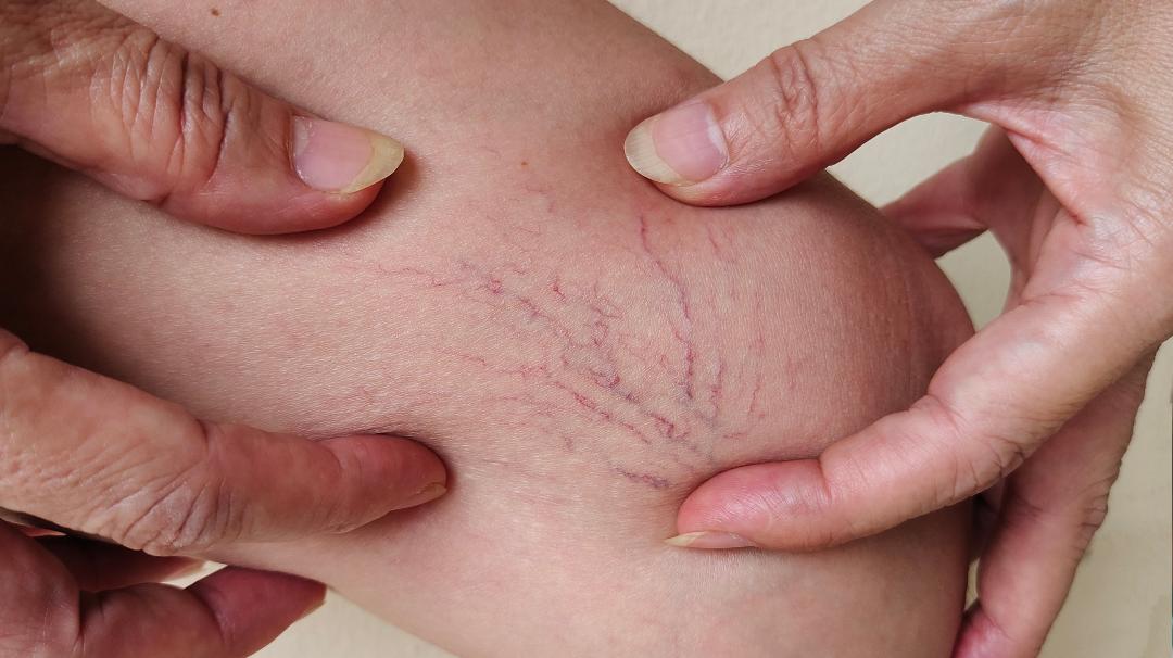

Telangiectasias or spider veins are dilated skin veins with a diameter of up to 1 mm. They often look like bluish or reddish lines in the shape of a spider’s web or a star.
Reticular veins are dilated veins located a little deeper and located at the skin’s transition to the subcutaneous tissue. They often form a branched network and are 1 to 3 millimeters in diameter.
Both types of veins most often appear on the outer side of the thigh, lower leg, and in the popliteal region (back of the knee), and generally have very little or no influence on venous hemodynamics.
In the case of reticular veins of the legs, symptoms such as a feeling of heavy legs, stabbing pain, or feeling of pressure in the legs may occur, while telangiectasias are asymptomatic.
It is important to distinguish reticular veins and telangiectasias (C1 stage according to the CEAP classification) in relation to varicose veins (C2 stage according to the CEAP classification).
The diagnosis is made during a thorough examination of the venous system, targeted questions (anamnesis), and clinical examination using Veinlite LEDX, a portable transilluminator specially designed to facilitate microsclerotherapy and ultrasound examination of veins. With an ultrasound examination, we rule out changes in the deep venous system and reflux of the sapheno-femoral confluence, which is located in the groin, or the sapheno-popliteal confluence, which is located in the popliteal fossa (pelvic region).
After establishing the diagnosis, together with the patient, we agree on a treatment plan, noting that the remodeling of the veins after treatment occurs within a few weeks and that several treatments are needed to achieve the desired effect.
Injecting a sclerosing agent (Aethoxysklerol) into the veins is called sclerotherapy, and sclerotherapy of reticular veins in telangiectasia is called microsclerotherapy (which is also the method of choice). Treatment of varicose veins with sclerotherapy is not recommended.
By injecting a sclerosing agent into the vein, the blood is squeezed out, and the agent then damages the vein endothelium (the inner layer of the vein wall) and thus leads to blockage of the vein that in the long run turn into a scar that can no longer conduct blood. Shortly after the initial loss of color of the veins, as a result of the injection of Aethoxysclerol, the treated area becomes slightly red, one of the signs of inflammation, which is the expected and normal effect of the sclerosant on the vein wall.
We use the sclerosing agent Aethoxysklerol with concentrations of 0.25%, 0.5%, or 1% in the form of liquid or foam depending on the change in the vein.
After the treatment, we apply local compression (band-aid or tampon), then an elastic compression stocking, which must be worn continuously 24 hours a day, and then for another two to seven days at least 8 hours a day, it is recommended to walk for 15 to 30 minutes immediately after the procedure. The only prohibitions after the treatment are the use of a sauna, solarium, or sunbathing, it is recommended to avoid prolonged sitting and standing, while other normal lifestyle habits are not prohibited.
Complications of the procedure are usually transient within a year, they are microthrombosis (dark blue or black spots), hyperpigmentation (darker skin at the treatment site), matting (the appearance of new small telangiectasias as a consequence of the reaction to the sclerosant) which occurs in less than 5% of treatments and is solved by laser treatment with the interruption of further sclerosing.
The first check-up is after one to two weeks, when possible microthrombosis is removed, while the second check-up is after six weeks, when it is possible to assess the final effect of the therapy.
Written by Dr. Frano Šimić, a specialist in vascular surgery